
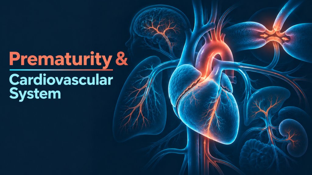
Factsheet Neonatal haemodynamics Physiology
The preterm heart isn’t a small term heart. It has less contractile machinery, a weaker grip on calcium and far less reserve, and on the first day it meets a sudden jump in afterload that it often can’t overcome.
In the first 24 hours, the danger for a very preterm baby is low blood flow rather than a low number on the monitor. Blood pressure is a poor guide to that flow, and the brain pays the price when flow drops and then recovers.
The immature myocardium
The difference starts at cellular level. The immature heart has fewer myofibrils per cross-sectional area, with a simpler internal layout, an underdeveloped sarcoplasmic reticulum and T-tubule system, altered calcium handling and different troponin expression, all of which mean less force per beat.1,2
Smaller, less mature cells
Preterm myocytes are smaller and less mature than term myocytes, with poorly arranged contractile filaments.2 The heart has a limited capacity to increase contractility on demand.1
Calcium from outside the cell
Because the sarcoplasmic reticulum is poorly developed and takes up calcium slowly, the preterm heart relies more on calcium entering across the cell membrane to contract.1,2
Little reserve
Put together, this gives a myocardium that copes poorly with a sudden rise in afterload and has little reserve when preload falls.2
Fewer β-receptors, less innervation
Contractility is further limited by fewer β-adrenergic receptors and less sympathetic innervation of the heart.1
Birth: an afterload shock
Clamping the cord removes the low-resistance placental bed, and systemic vascular resistance rises straight away.1 A term heart takes this in its stride. The immature preterm myocardium often can’t, and systemic blood flow falls.2,3
In a cohort of 126 babies born before 30 weeks, low SVC flow was common in the first day and linked to lower gestation, higher upper-body vascular resistance, larger ductal shunts and higher mean airway pressure.3 A second cohort found low flow in 34% of babies, peaking at 5–10 hours (31%) and almost gone by 24 hours (3%).4
Receptors and the drugs we reach for
Adrenergic receptors are still maturing in preterm babies, and receptor polymorphisms and differences in drug handling all affect how an inotrope behaves.12 Endogenous catecholamine levels may already be high after birth.12
This shows up at the bedside. In a randomised trial in preterm babies with low SVC flow, dopamine raised blood pressure more, but dobutamine raised SVC flow more, and 40% of babies didn’t respond to either drug.13 A pressure rise bought with vasoconstriction can mean more afterload for a heart that is already struggling. (Expert consensus: a response skewed towards peripheral vasoconstriction rather than cardiac output in the most immature babies is a widely taught explanation, but the receptor data behind it weren’t confirmed in this review.)
Evidence that dopamine or dobutamine actually improves outcomes in preterm babies remains limited.11
The ductus arteriosus
Ductal closure is driven by rising oxygen, falling prostaglandin levels, endothelin and catecholamines.1 The vascular responses behind closure depend on gestational age, which is one reason the duct stays open longer in preterm babies.6 Functional closure usually happens within hours of birth, while anatomical closure can take weeks to months, especially in preterm infants.1
Blood leaves the systemic circulation
As pulmonary vascular resistance falls, left-to-right ductal shunting increases, flooding the lungs and taking blood away from the body.6 Larger ductal shunts were linked to low SVC flow on day one.3
Organs downstream
Substantial shunting may increase the risk of intraventricular haemorrhage, necrotising enterocolitis and death.6 (Expert consensus: diastolic “run-off” lowers diastolic pressure and post-ductal perfusion.)
The brain: when flow fails, then returns
Developmental or acquired problems with cerebral autoregulation contribute to preterm brain injury, and repeated episodes of ischaemia and reperfusion are a common pathway to both IVH and white matter injury.7 When cerebral blood flow is pressure-passive, low pressure risks ischaemia and pressure surges risk capillary rupture and haemorrhage.8
In Kluckow and Evans’ cohort, late IVH was first seen after SVC flow had improved, and the grade of IVH tracked the severity and duration of the low-flow period.3 Low flow on day one was also linked to death or disability at 3 years.5 Watch for low flow early, and handle its recovery gently.
Hormones and adrenal reserve
Relative adrenal insufficiency, meaning a cortisol response that is too small for the degree of illness, is increasingly recognised as a cause of haemodynamic instability and hypotension in sick newborns, though data are still limited.10 It may come with down-regulation of adrenergic receptors, which helps explain why some babies stop responding to catecholamines.9
(Expert consensus: HPA-axis suppression and immature adrenal steroid synthesis are commonly cited mechanisms, and cardiovascular hormone levels are thought to rise with gestation. These points weren’t verified against primary sources for this factsheet.)
What this means at the cot side
Don’t
References
- Vrancken SL, van Heijst AF, de Boode WP. Neonatal hemodynamics: from developmental physiology to comprehensive monitoring. Frontiers in Pediatrics. 2018;6:87. doi:10.3389/fped.2018.00087
- Ping P, Yu B, Xu R. Monitoring and evaluation of hypotension in the extremely preterm. Frontiers in Cardiovascular Medicine. 2024;11:1477337. doi:10.3389/fcvm.2024.1477337
- Kluckow M, Evans N. Low superior vena cava flow and intraventricular haemorrhage in preterm infants. Archives of Disease in Childhood – Fetal and Neonatal Edition. 2000;82(3):F188–94. doi:10.1136/fn.82.3.f188
- Osborn DA, Evans N, Kluckow M. Clinical detection of low upper body blood flow in very premature infants using blood pressure, capillary refill time, and central-peripheral temperature difference. Archives of Disease in Childhood – Fetal and Neonatal Edition. 2004;89(2):F168–73. doi:10.1136/adc.2002.023796
- Hunt RW, Evans N, Rieger I, Kluckow M. Low superior vena cava flow and neurodevelopment at 3 years in very preterm infants. Journal of Pediatrics. 2004;145(5):588–92. doi:10.1016/j.jpeds.2004.06.056
- Hamrick SEG, Hansmann G. Patent ductus arteriosus of the preterm infant. Pediatrics. 2010;125(5):1020–30. doi:10.1542/peds.2009-3506
- Vesoulis ZA, Mathur AM. Cerebral autoregulation, brain injury, and the transitioning premature infant. Frontiers in Pediatrics. 2017;5:64. doi:10.3389/fped.2017.00064
- Lou HC. Perinatal hypoxic-ischemic brain damage and intraventricular hemorrhage: a pathogenetic model. Archives of Neurology. 1980;37(9):585–7. doi:10.1001/archneur.1980.00500580081017
- Noori S, Seri I. Pathophysiology of newborn hypotension outside the transitional period. Early Human Development. 2005;81(5):399–404. doi:10.1016/j.earlhumdev.2005.03.007
- Fernandez EF, Watterberg KL. Relative adrenal insufficiency in the preterm and term infant. Journal of Perinatology. 2009;29 Suppl 2:S44–9. doi:10.1038/jp.2009.24
- Lingwood BE, Eiby YA, Bjorkman ST, Miller SM, Wright IMR. Supporting preterm cardiovascular function. Clinical and Experimental Pharmacology and Physiology. 2019;46(3):274–9. doi:10.1111/1440-1681.13044
- Garvey AA, Kooi EMW, Dempsey EM. Inotropes for preterm infants: 50 years on are we any wiser? Frontiers in Pediatrics. 2018;6:88. doi:10.3389/fped.2018.00088
- Osborn D, Evans N, Kluckow M. Randomized trial of dobutamine versus dopamine in preterm infants with low systemic blood flow. Journal of Pediatrics. 2002;140(2):183–91. doi:10.1067/mpd.2002.120834